Seeing the Pelvic Floor in Real Time: How Rehabilitative Ultrasound Is Changing Physical Therapy
Pelvic floor physical therapy is changing in exciting ways. One of the biggest advances is the use of Rehabilitative Ultrasound Imaging (RUSI). This tool helps therapists and patients better understand how the deep core muscles work together. These muscles include the pelvic floor and the abdominal wall, especially a deep muscle called the transversus abdominis (TrA). When these muscles work well, they support the spine, control pressure in the abdomen, and help with everyday movements like lifting, walking, and reaching. When they do not work well, people may have pain, weakness, or trouble with bladder and bowel control.
RUSI has two main roles in rehab. First, it serves as a tool to assess muscles. Therapists can see the size and shape of muscles and how they change during contraction. Second, and just as important, it acts as a biofeedback tool. This means patients can see their muscles working in real time on a screen. Instead of guessing if they are doing an exercise correctly, they get clear visual feedback. This makes learning faster and more effective.
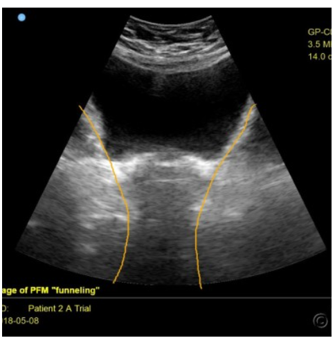
Photo courtesy of Ramona Horton, DPT
Many people struggle to activate their deep core muscles correctly. They may overuse larger, more superficial muscles instead. This is where RUSI shines. Research shows that visual feedback from ultrasound is more effective than just verbal cues or touch. When patients can see their muscles, they are better able to perform motor control exercises. These exercises focus on gentle, precise muscle activation rather than big, forceful movements.
One key exercise in pelvic floor and abdominal rehab is abdominal activation, sometimes called “hollowing” in fitness classes. This exercise targets the transversus abdominis. Studies show that when people use ultrasound feedback during this exercise, they activate the TrA more selectively. In simple terms, they use the right muscle at the right time. This is important because proper muscle coordination helps protect the spine and pelvic organs.
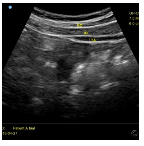
Photo Courtesy of Ramona Horton, DPT
Another benefit of RUSI is improved endurance. It is not just about turning a muscle on—it is about keeping it on. With ultrasound feedback, patients can better maintain a contraction over time. For example, during a 30-second hold, people using RUSI show stronger and more consistent muscle activity. This matters because daily activities often require low-level muscle activation over longer periods, not just short bursts of effort.
As patients improve, therapy does not stay on the treatment table. A key goal is to transfer these skills into real life. Research shows that training the TrA in a lying position with ultrasound feedback can carry over to standing tasks. These include lifting, reaching, and other functional movements. Even better, these improvements can last for months after training ends. This supports a step-by-step approach: start with simple positions, then progress to more complex, weight-bearing tasks.
Muscle function changes depending on the task. For example, how the TrA works during standing or a single-leg squat is different from how it works lying down. Because of this, newer methods look at functional activation ratios. This means comparing muscle activity during real tasks instead of just at rest. This gives a more accurate picture of how the body works in daily life.
RUSI is also helpful for people with pain, especially low back pain. Studies show that individuals with ongoing back pain often have reduced activation of the transversus abdominis during standing tasks. By using ultrasound during these activities, therapists can identify these deficits and guide treatment more effectively. This helps patients retrain their muscles in a way that directly relates to their symptoms.
Another important area is post-surgical rehabilitation. After procedures like hernia repair or abdominal wall reconstruction, proper muscle function is critical. Research shows that structured rehab programs can reduce the risk of complications, such as hernia recurrence or abdominal bulging. Strengthening the abdominal wall and improving coordination with the pelvic floor are key parts of recovery. RUSI can play a role here by guiding safe and effective muscle activation during the healing process.
While RUSI is a powerful tool, it is important to understand its limits. Changes in muscle thickness seen on ultrasound do not always equal muscle strength or activity. The relationship is strongest during low-level, controlled contractions. Factors like body position and effort level can affect the readings. This means therapists must use RUSI along with clinical judgment and other assessments.
In pelvic floor physical therapy, the connection between the abdominal wall and pelvic floor is essential. These muscles work as a team to manage pressure, support organs, and stabilize the body. When one part is not working well, the whole system can be affected. By combining RUSI with functional training, therapists can help patients rebuild this coordination step by step.
In practice, a typical program might start with simple breathing and gentle activation exercises in a lying position. Using ultrasound, the patient learns how to engage the deep core without overusing other muscles. As control improves, exercises progress to sitting, standing, and eventually more dynamic tasks like lifting or squatting. Throughout this process, the focus remains on quality of movement, not just quantity.
This approach fits well with modern rehabilitation principles. It is patient-centered, evidence-based, and focused on real-life function. Instead of just treating symptoms, it addresses the underlying movement patterns that contribute to pain or dysfunction.
In summary, rehabilitative ultrasound imaging is a valuable tool in pelvic floor and abdominal wall rehabilitation. It helps assess muscle function, improves exercise performance, and supports the transfer of skills to daily activities. For patients, this means clearer guidance, better outcomes, and a more active role in their own recovery.
Disclaimer: This blog is here for your help. It is the opinion of a Licensed Physical Therapist. If you experience the symptoms addressed you should seek the help of a medical professional who can diagnose and develop a treatment plan that is individualized for you.
Related Posts

Breathe your way to a healthy pelvic floor: Diaphragmatic Breathing 101
It is a continuous conversation in my treatment room with my clients – every single patient, every day hears something…

The Real Reason Golfers Lose Power, Distance, and Comfort on the Course
Golf may look smooth and effortless, but the body works extremely hard during every swing. A powerful golf swing depends…
Constipation and GLP-1 Medications: What You Need to Know Before Symptoms Start
Constipation is a common but often overlooked side effect in people taking GLP-1 receptor agonists, a class of medications widely…