The Psoas Muscle: the forth horseman in pelvic and spinal stability.
The psoas muscle gets a lot of press for being overactive or tight. Everyone wants to release their psoas. It seems to stalk my Instagram feed.
So, what does the psoas really do and is it truly tight on everyone? Or is the psoas muscle’s tightness indicative of weakness somewhere else?
The psoas muscle is the bridge between the lumbar spine, the pelvis and the leg. At the top, the psoas attaches to the spine in 3 places: through the arcuate ligament which has a direct attachment to the diaphragm, and the 1st, 2nd, and 3rd lumbar vertebrae of the spine. In some texts, it appears that the diaphragm and the ligament blend with anterior ligaments of the spine. The psoas also attaches to the spine on the transverse processes from L1-L5 and on the discs of T12-L1 to L4-L5. Whew! If your head is spinning here is an image that might help clear it up!
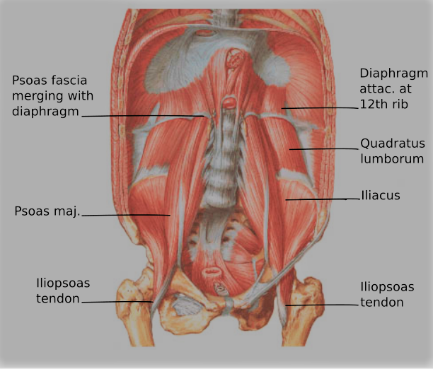
At the bottom, the psoas fascia (connective tissue) attaches firmly on the pelvis forming a ligament that attaches the muscle firmly onto the body of the pelvis itself. As the psoas muscle travels down from the lumbar spine and into the pelvis, the fascia blends into the pelvic floor muscle and fascia. The psoas tendon then combines with the iliacus muscle tendon and becomes the iliopsoas, attaching to the femur (thigh bone).
As you can see from the anatomical attachments of the iliacus and psoas, this muscle is a flexor of the hip and has an even greater role in Lumbar spine (low back) stabilization. Because the psoas has fascial connections to the diaphragm and pelvic floor, the psoas acts as a link in abdominal pressure and stability. The psoas plays a vital role in lumbar spine stabilization by increasing compression forces on the vertebral discs. This compression helps to improve stability at individual vertebral segments and reduce sheer across vertebrae.
Because of the fascial connections between the pelvic floor and the iliopsoas, the iliopsoas can be considered as part of the deep core—similar to the transverse abdominus, diaphragm and multifidus muscles. The iliopsoas muscle also crosses in front of the SI joint and can help to create force and stability at the SI joint and contribute to maintaining intra-abdominal pressure. As discussed in other blogs, the pelvic floor contracts and coordinates with the transverse abdominus, diaphragm, and the multifidus. If one of these muscles becomes weakened or there is a decrease in the coordination of the muscle activations, there can be a decrease in tension between the vertebra, leading to potential injury or pain.
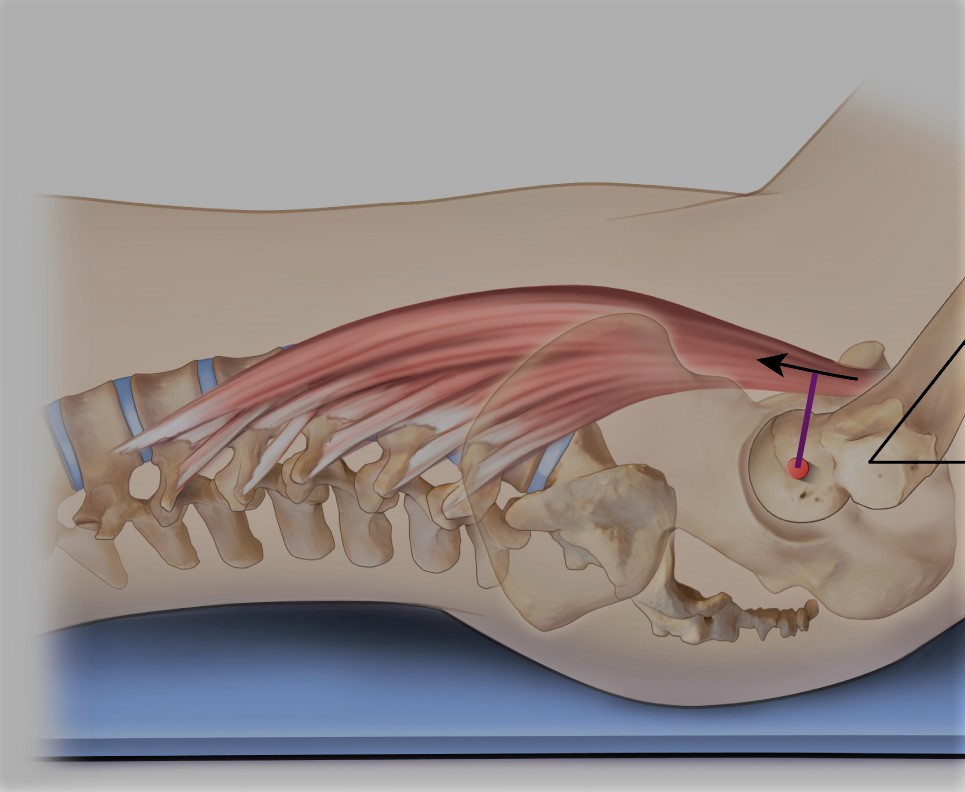
As you can see from the way the iliopsoas travels over the hip joint to attach at the femur, it creates a functional wall in front of the hip. This allows it to assist in the centering the femoral head in the acetabulum of the hip. To make it simple: it helps keep the ball in the socket of the joint. This decreases anterior femoral head (the ball) from shifting forward and decrease the strain on the anterior labrum (the joint capsule tissues) improving hip health.
The psoas is a complex muscle with multiple roles in the spine, abdominal wall, and hip. Keeping this in mind, if psoas keeps needing to be released, a different muscle in the system is most likely not doing its job. This can be a strength or a coordination issue. This is where PT comes in. We can do a functional movement analysis and see where the system is not as coordinated so that the psoas does not work overtime for a muscle that needs to step up.
Disclaimer: This blog is here for your help. It is the opinion of a Licensed Physical Therapist. If you experience the symptoms addressed you should seek the help of a medical professional who can diagnose and develop a treatment plan that is individualized for you.
Related Posts

The Real Reason Golfers Lose Power, Distance, and Comfort on the Course
Golf may look smooth and effortless, but the body works extremely hard during every swing. A powerful golf swing depends…
Seeing the Pelvic Floor in Real Time: How Rehabilitative Ultrasound Is Changing Physical Therapy
Pelvic floor physical therapy is changing in exciting ways. One of the biggest advances is the use of Rehabilitative Ultrasound…

Constipation and GLP-1 Medications: What You Need to Know Before Symptoms Start
Constipation is a common but often overlooked side effect in people taking GLP-1 receptor agonists, a class of medications widely…